Dermatological peptides in research: GHK-Cu, Matrixyl, Argireline and the science of cosmeceuticals
Skin, the model laboratory for peptides
Skin constitutes a preferred research terrain for peptide biology: accessible tissue, well-characterised stratified structure (epidermis, dermis, hypodermis), measurable turnover, biological response quantifiable by histology, biochemistry, confocal imaging. Preclinical studies have documented the action of several peptide families on extracellular matrix, cutaneous angiogenesis, pigmentation, tissue regeneration and ageing.
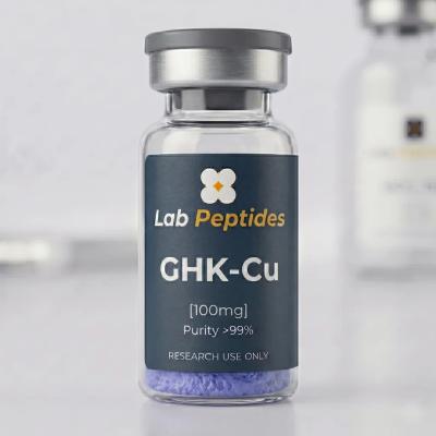
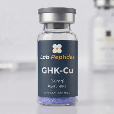
This dossier synthesises available scientific data on the main dermatological peptides used in cosmetic and medical research: GHK-Cu, Matrixyl (palmitoyl pentapeptide), Argireline (acetyl hexapeptide), Copper Tripeptide-1, Palmitoyl tripeptides. Absolute reminder: the peptides sold by Lab Peptides France are strictly intended for in vitro research and preclinical RUO models. No human cosmetic application, no ready-to-use topical formulation, no medical indication is covered by our products.
GHK-Cu: the historical copper tripeptide
Structure and complexation
GHK (Glycyl-L-Histidyl-L-Lysine) is an endogenous tripeptide identified by Loren Pickart in 1973 in human plasma. Its plasma concentration declines with age (~200 ng/mL at 20 years, ~80 ng/mL at 60 years), an observation that has fed the hypothesis of involvement in tissue ageing. GHK spontaneously forms a 1:1 complex with copper(II) via coordination by the histidine imidazole nitrogen, the glycine α-amino nitrogen, and carboxylate oxygens. The GHK-Cu complex (peptide alone molecular mass: 340.4 Da) is the biologically active form in the majority of published studies.
Documented biological mechanisms
More than 100 preclinical studies have documented various GHK-Cu effects, mainly on cultured fibroblasts, skin explants and murine wound healing models:
- Stimulation of type I and III collagen synthesis in cutaneous fibroblasts
- Increased elastin and modulation of metalloproteinases (MMP-2, MMP-9) — anti-degradation profile
- Activation of decorin and glycosaminoglycan synthesis (hyaluronic acid, chondroitin sulfate)
- VEGF induction and angiogenesis documented on chorioallantoic membrane and in vivo
- Anti-oxidant activity by modulating expression of SOD, catalase, glutathione peroxidase
- Broad transcriptomic modulation: microarray studies have shown more than 4000 genes modulated by GHK-Cu on fibroblasts, with a tissue rejuvenation signature
Standard experimental models
- Primary fibroblast cultures human (Hs27, BJ) and murine, concentrations 10-100 µg/mL GHK-Cu
- Ex vivo skin explant models to test penetration and dermal matrix action
- Murine wound models (excisional wound, burn) with kinetic closure measurement and histology
- Full-thickness healing models with immunohistochemical staining (collagen I, III, α-SMA for myofibroblasts)
Matrixyl: the synthetic palmitoyl pentapeptide
Structure
Matrixyl (Pal-KTTKS, generic name palmitoyl pentapeptide-4) is a synthetic pentapeptide Lys-Thr-Thr-Lys-Ser derived from the C-terminal propeptide of type I procollagen. A palmitic (C16) chain is added at the N-terminus to improve lipophilicity and cutaneous penetration. Molecular mass: 802.1 Da.
Biological rationale
The KTTKS fragment is recognised as a negative feedback signal of collagen synthesis: during C-terminal propeptide cleavage, it accumulates locally and signals the fibroblast that enough collagen is produced. By exogenously applying Pal-KTTKS at low concentration to fibroblasts, the observed effect is paradoxically inverse: stimulation of type I and III collagen production and fibronectin. Robinson et al. studies (2005) are the historical reference, with multiple confirmations since.
Palmitoyl tripeptide-5 and derivatives
Several derivatives have been developed in the 2000s-2010s:
- Palmitoyl tripeptide-1 (Pal-GHK): combination of native GHK with a palmitic chain for improved topical penetration
- Palmitoyl tripeptide-5 (Pal-KVK): TGF-β growth factor mimetic, documented as collagen inducer
- Palmitoyl tetrapeptide-7 (Pal-GQPR): studied for cutaneous anti-inflammatory effect
These variants all exploit the same principle: short signal peptide + lipid chain for improved membrane penetration while preserving biological activity.
Argireline: the "topical Botox" hexapeptide
Structure and mechanism
Argireline (acetyl hexapeptide-3 or acetyl hexapeptide-8, depending on nomenclature) is a hexapeptide with sequence Ac-Glu-Glu-Met-Gln-Arg-Arg-NH₂ (EEMQRR), mimetic of the N-terminal site of SNAP-25. Molecular mass: 888.1 Da. Its proposed mechanism is interference with assembly of the SNARE complex necessary for synaptic exocytosis, particularly at the neuromuscular junction.
Mechanistic rationale
Botulinum toxin A cleaves SNAP-25 at a specific site, paralysing cholinergic transmission. Argireline is presented as a competitive inhibitor of the SNARE complex: by mimicking a portion of SNAP-25, it would prevent productive SNARE complex assembly, thus reducing neurotransmitter release. The documented preclinical effect on PC12 cells (pheochromocytoma) shows a reduction in calcium-dependent catecholamine release.
Limitations and controversies
Two questions deserve critical examination:
- Cutaneous penetration: Argireline is a polar hexameric peptide (MW ~880 Da, net charge); its permeation through intact stratum corneum is biophysically limited. Franz cell diffusion studies show real but low penetration, which depends heavily on the vehicle (liposomes, nanoemulsions).
- Effective in vivo activity: published clinical studies report modest effects on expression wrinkle depth. Comparison with injectable botulinum toxin remains a mechanistic chasm.
In RUO research, Argireline remains an interesting tool for studying the SNARE mechanism on cellular models, independently of the cosmetic claims surrounding it.
Molecular mechanisms of cutaneous ageing
To interpret the effects of dermatological peptides, the molecular alterations of cutaneous ageing must be understood:
Intrinsic ageing (chronological)
- Decreased collagen synthesis by fibroblasts (–1% per year after 30)
- AGE accumulation (Advanced Glycation End-products) by non-enzymatic glycation
- Fibroblastic senescence with SASP (Senescence-Associated Secretory Phenotype), chronic pro-inflammatory secretion
- Dermal atrophy, reduced vascularisation, loss of subcutaneous adipocytes
Extrinsic ageing (photoageing)
- UV-B damage: cyclobutane pyrimidine dimers, keratinocyte DNA alteration
- UV-A and oxidative stress: ROS, lipid peroxidation, protein carbonylation
- MMP overexpression (MMP-1, MMP-3, MMP-9) via AP-1 activation, degrading collagen and elastin
- Solar elastosis: accumulation of degraded and dystrophic elastin in the dermis
Dermatological peptides act at different levels of these cascades: anabolic stimulation (GHK-Cu, Matrixyl), catabolic inhibition (MMP modulation), anti-oxidant protection (GHK-Cu), inflammatory signalling modulation.
Analysis and quality control of dermatological peptides
Dermatological peptides share general analytical requirements with one specificity: formulation stability is a central issue.
- MS identity: ESI-LRMS or HRMS, verification of modifications (palmitoylation, acetylation, cuprification). For GHK-Cu, UV-Vis spectrum at 540-560 nm confirms copper complexation
- HPLC purity: ≥ 98% in reverse phase. For palmitoylated peptides (Matrixyl, Pal-GHK), C18 column with acetonitrile gradient adapted to lipophilicity
- Copper content (GHK-Cu only): ICP-MS or AA spectroscopy. Target Cu:peptide molar ratio = 1:1. A ratio > 1 suggests free copper, pro-oxidant and potentially cytotoxic
- Chemical stability: oxidation (methionines for Argireline), isomerisation, hydrolysis of the palmitic bond
- Formulation stability: accelerated test 40°C/75% RH for aqueous and oily formulations. Lipopeptides can form self-assemblies (micelles, vesicles) that affect bioavailability
Study pathways: in vitro, ex vivo, in vivo
Cell cultures
- Primary fibroblasts (neonatal, adult, senescent) on 2D collagen-coated or 3D scaffolds
- Keratinocytes HaCaT or primary for barrier and pigmentation studies (melanocyte co-culture)
- Skin equivalent models: EpiSkin, Phenion, reticulated skin (raft cultures)
Ex vivo
- Human skin explants (plastic surgery) maintained in interface culture 7-14 days
- Organotypic perfusion of porcine skin, frequent model for penetration studies
In vivo
- Murine wound models: 6 mm dorsal excisional wound, kinetic closure measurement
- Photoageing: SKH-1 hairless under chronic UV-B, wrinkle quantification by optical profilometry
- Diabetes (db/db): delayed healing, efficacy model for GHK-Cu
- Porcine models: skin close to human, used for clinical translation studies
Research dermatological peptides FAQ
Why is a native tripeptide (GHK) active?
GHK receptors are not yet all identified, but recognition seems to involve peptide transporters (PepT1/PepT2) and direct interactions with DNA and transcription factors in the nucleus after penetration. Copper presence is essential for redox activity and for stabilising the active conformation.
GHK-Cu vs GHK without copper: what difference?
Free GHK has residual biological activity, but the majority of documented effects require the Cu(II) complex. In solution, GHK chelates physiologically present copper (albumin, ceruloplasmin); in pure formulation, copper must be added exogenously to ensure activity.
Matrixyl vs Pal-GHK: which to choose in research?
It depends on the scientific question. Matrixyl targets collagen synthesis feedback via a propeptide mimetic; Pal-GHK combines GHK action and topical penetration. For comparative lipid penetration studies, both are reference compounds.
Is copper cytotoxic?
Free copper (Cu²⁺ ionic) is pro-oxidant and cytotoxic beyond 50-100 µM. GHK-Cu is non-toxic up to high concentrations (> 500 µM in most cell lines), because copper is sequestered in a coordinated geometry that prevents it from catalysing Fenton reactions. Cu:peptide ratio control is critical to guarantee this innocuity.
Do dermatological peptides penetrate the stratum corneum?
Most hydrophilic peptides (GHK, Argireline) have intrinsically low penetration. Enhancement approaches are: palmitoylated peptides (Matrixyl, Pal-GHK), liposomes, microemulsions, polymer nanoparticles, microneedles, iontophoresis. In cellular research, penetration is not a problem (direct application), but it becomes critical for any topical extrapolation.
Conclusion
Dermatological peptides constitute a scientifically rich family: GHK-Cu as endogenous regulatory molecule, Matrixyl as feedback mimetic, Argireline as SNARE inhibitor. Each illustrates a different intervention strategy on cutaneous biology. In rigorous research, requirements are: complete analytical control (HPLC ≥ 98%, MS identity, Cu ratio for GHK-Cu, formulation stability), adapted experimental models (primary fibroblasts, explants, in vivo photoageing or wound models), exhaustive documentation for publication. RUO reminder: no human cosmetic use, no ready-to-use formulation, no topical efficacy claim. These peptides are research tools — their evaluation in cosmetic or medical formulation falls under a distinct regulatory framework (medical devices, cosmetics, medicines).
Cutaneous antimicrobial peptides and innate defence
Beyond cosmetic anti-ageing peptides, skin is also the stage of a peptide innate defence that is the subject of active research. Cutaneous antimicrobial peptides (AMPs) include defensins (β-defensins hBD-1, hBD-2, hBD-3), cathelicidin LL-37, psoriasin (S100A7), RNase 7. Produced by keratinocytes and resident immune cells, they constitute the first line of defence against bacteria, fungi and viruses.
AMP research exploits their:
- Broad-spectrum bactericidal activity via membrane perturbation (colloido-osmotic mechanism, pore formation)
- Low resistance to bacterial selection (physical mechanism, little subject to mutation)
- Potential as antibiotic substitute in multi-resistance context
- Immunomodulatory activity: LL-37 stimulates leukocyte migration, neovascularisation, reepithelialisation
In preclinical research, new-generation synthetic antimicrobial peptides (derivatives of magainin, indolicidin, protegrins) are tested on infected wound models. Compounds like pexiganan (magainin analogue), LL-37 and fragments (KR-12, KS-30) are studied for their action in Pseudomonas aeruginosa cutaneous biofilm and infected diabetic wounds.
Pigmentation and peptides: melanogenesis, tyrosinase and new modulators
Biological pathways of melanogenesis
Melanogenesis involves an enzymatic cascade in melanocytes: tyrosine → DOPA → DOPAquinone → eumelanin/pheomelanin. Key enzymes are tyrosinase, TRP-1 (tyrosinase-related protein 1) and DCT/TRP-2. Transcriptional regulation passes through MITF (Microphthalmia-Associated Transcription Factor), itself modulated by the α-MSH / MC1R pathway (melanocortin receptor type 1).
Peptides modulating pigmentation
- α-MSH (Melanocortin, 13 aa): melanogenesis-stimulating hormone, reference endogenous peptide. Its analogue afamelanotide (Melanotan-I) and Melanotan-II are used in pigmentation and photoprotection research. RUO caution: these compounds are sold strictly for research and have no validated cosmetic indication in humans. Their recreational uses reported in the grey literature are outside the scientific framework.
- Oligopeptide-51 and -55: anti-hyperpigmentation peptides, studied for solar spots and senile lentigines
- Nonapeptide-1: α-MSH antagonist, melanogenesis inhibitor in cosmetic research
- Agouti signaling protein (ASIP): physiological MC1R antagonist, oriented toward pheomelanin rather than eumelanin
Experimental pigmentation models
Studies classically use: murine B16 melanocytes (F10 line, F1 line for differentiation), primary human melanocytes (NHEM), melanocyte-keratinocyte co-cultures, pigmented skin equivalents (MelanoDerm, Phenion FT). Measurements: colorimetric melanin dosing (absorbance 405-490 nm), tyrosinase activity by L-DOPA oxidation, MITF expression by qPCR, melanosomal translocation by microscopy.
Topical formulation and advanced delivery systems
Cutaneous penetration remains the main bottleneck for dermatological peptides. Several formulation strategies have been developed:
Liposomes and transfersomes
Classical liposomes (phosphatidylcholine + cholesterol) encapsulate hydrophilic peptides and facilitate transit through the stratum corneum by membrane fusion/fission. Transfersomes (ultradeformable liposomes with edge activators like Tween-80) cross pores well below their diameter by elastic deformation.
Polymer nanoparticles
PLGA (poly-lactide-co-glycolide), chitosan, functionalised polysaccharides: peptide encapsulation, controlled release over several hours/days. Used for Matrixyl, Argireline, GHK-Cu with demonstrations of improved cutaneous bioavailability.
Microemulsions and nanoemulsions
Oil-water-surfactant mixture thermodynamically (microemulsion) or kinetically (nanoemulsion) stable. Droplets 10-200 nm. Improved penetration by transient destructuring of the stratum corneum.
Soluble microneedles and patches
Recent technology (2010s): polymer needle matrices (dextran, hyaluronic acid, PVA) loaded with peptide. Painless insertion into the epidermis, progressive dissolution. Allows intradermal delivery for high MW peptides or to reach deep dermal targets.
Iontophoresis and sonophoresis
Iontophoresis: application of a low electric current to increase penetration of charged peptides. Sonophoresis: low-frequency ultrasounds to temporarily permeabilise the stratum corneum. Techniques used in comparative research to evaluate maximum achievable bioavailability.
Reference clinical studies and methodological limitations
In cosmetic dermatology, clinical studies on peptides are often methodologically limited: reduced sample sizes (n = 20-50), short duration (4-12 weeks), subjective measurements (self-evaluation, photography), absence of strict vehicle control. For researchers wishing to compare their preclinical results to clinical literature, these limitations are important to know:
- Robinson et al. 2005: pivotal Matrixyl (Pal-KTTKS) study, double-blind vehicle-controlled, n = 93, 12 weeks. Significant reduction in wrinkles and skin roughness measured by profilometry. Quality methodological reference.
- Pickart 2008, 2015: narrative reviews on GHK-Cu, founded on a compilation of in vitro and animal studies with few randomised clinical trials.
- Blanes-Mira et al. 2002: original Argireline study, n = 10, topical 2×/day for 30 days. Declared 27% reduction in wrinkle depth. Small sample, open design, results to interpret with caution.
For a research laboratory, these studies offer benchmarks but do not constitute a rigorous level A evidence framework. Ambitious preclinical protocols should aim to reproduce underlying mechanistic hypotheses, not to replicate criticisable clinical designs.
Emerging peptides in research dermatology
Thymosin β-4 in cutaneous healing
Beyond TB-500 (acetyl-lys-lys-lys-thymosin β-4 form), native Thymosin β-4 is studied in diabetic cutaneous healing. Its effects: keratinocyte migration stimulation, angiogenesis, anti-inflammation. Potential applications: chronic wounds, venous ulcers, diabetic foot wounds.
FGF-2 peptide mimetics
Small peptide molecules mimicking Fibroblast Growth Factor 2 (FGF-2) for regeneration stimulation without systemic side effects of the complete growth factor. Interest in radiation wounds, hypertrophic scars.
MMP inhibitor peptides
Anti-degradation approach: TIMPs (Tissue Inhibitor of Metalloproteinases) mimetic peptides or peptides selectively inhibiting MMP-1, MMP-8, MMP-9. Research on cutaneous ageing, chronic ulcerations.
Circular peptides and cyclotides
Structure stabilised by disulfide bridge, considerably increased proteolytic resistance. Research on cyclotidic scaffolds to deliver dermatological functions with prolonged topical stability.
Molecular chaperone peptides
αB-crystallin (HSPB5) and its peptide derivatives, studied to protect dermal proteins against aggregation and oxidative damage in photoageing.
Regulation and boundary between cosmetic, medical device and medicine
In Europe, topical peptides fall under three distinct regulatory frameworks according to their claim and mechanism:
Cosmetic (EC Regulation 1223/2009)
- Surface cutaneous action, beauty and hygiene claims
- No therapeutic claim authorised
- Product information file (PIF), safety evaluation
- Examples: Matrixyl, Argireline, Pal-GHK in anti-ageing serums
Medical device (EU Regulation 2017/745)
- Main physical/mechanical action, medical claim
- Classes I, IIa, IIb, III according to risk
- CE marking mandatory, notified bodies for class IIa+
- Example: GHK-Cu dressings for wound healing
Medicine (Directive 2001/83/EC)
- Explicit therapeutic claim, pharmacological action
- MA mandatory (EMA or national agencies), phase I-III clinical trials
- Example: Afamelanotide for erythropoietic protoporphyria (MA 2014)
This segmentation has direct implications for research: a molecule can change regulatory status according to its final formulation and claims. An academic research project must clarify from the outset which framework it is destined for, which conditions the nature of data to generate (regulatory toxicology, controlled trials, pharmacovigilance).
Perspectives: artificial intelligence and dermatological peptide design
Generative AI tools (AlphaFold 3, RFdiffusion, ESM-3) are transforming the design of new dermatological peptides:
- De novo design of peptides binding to specific cutaneous receptors (EGFR, FGFR, MC1R)
- Cutaneous penetration prediction by QSAR/ML models integrating physicochemical properties
- Multiparametric optimisation: stability, solubility, activity, non-toxicity treated simultaneously
- Virtual peptide libraries computationally screened before real SPPS synthesis
For an academic laboratory, these approaches reduce the number of peptides to physically synthesise by several orders of magnitude. The combination of AI + high-throughput biology (high-density screens on fibroblasts or keratinocytes) becomes the dominant paradigm for 2nd and 3rd generation dermatological peptide discovery. RUO reminder: regardless of the level of technological sophistication, the final destination of peptides sold by Lab Peptides France remains laboratory research, and no ready-to-use cosmetic application or clinical claim is covered.