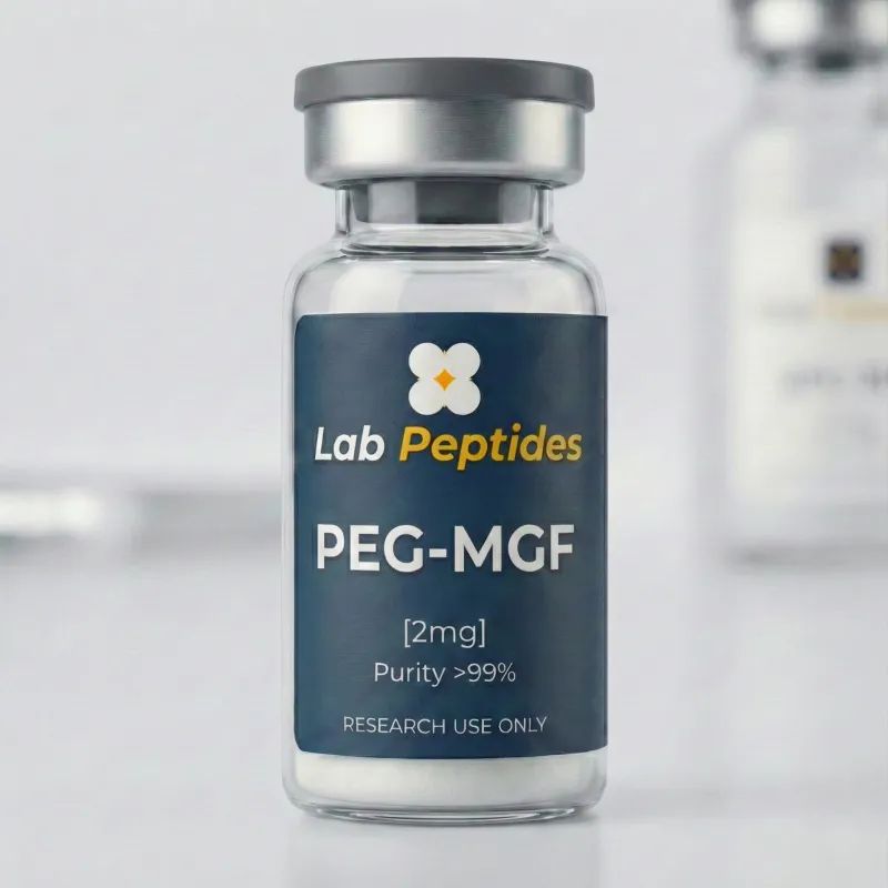
PEG-MGF 2mg Performance
Pegylated variant of Mechano Growth Factor. Muscular IGF-1 isoform studied for post-exercise muscle fiber regeneration.
Targeted post-effort muscle recovery. Pegylated Mechano Growth Factor for extended 24-48h action. Directly stimulates muscle satellite cells — boosted hypertrophy and recovery. The post-training injection that changes everything for advanced athletes.

Product information
PEG-MGF 2mg is a pegylated stabilized version of Mechano Growth Factor, muscle-specific splice variant of IGF-1 identified as IGF-1Ec. Discovered by Geoffrey Goldspink's team at the Royal Free Hospital in London in the late 1990s, this peptide represents the endogenous growth factor responsible for activating muscle satellite cells in response to mechanical stress and intense exercise.
The native MGF molecule presents an additional E-domain sequence of 24 amino acids at the C-terminus (Tyr-Gln-Pro-Pro-Ser-Thr-Asn-Lys-Asn-Thr-Lys-Ser-Gln-Arg-Arg-Lys-Gly-Ser-Thr-Phe-Glu-Glu-Arg-Lys) absent from hepatic systemic IGF-1. This unique sequence is generated by alternative splicing specific to muscle tissue after mechanical damage, and carries the mitogenic biological activity on Pax7+ satellite cells. The half-life of native MGF is however extremely short (less than 5 minutes in vivo) due to rapid peptidase degradation.
Pegylation (covalent attachment of a polyethylene glycol chain of approximately 20 kDa on the base molecule) dramatically extends plasma half-life to 5-7 days, transforming a local pulsatile molecule into a systemic long-acting agent. Atlas Lab formulates PEG-MGF 2mg as high-purity lyophilizate (>98% HPLC) for research applications exclusively. Strict RUO — not intended for human, veterinary, diagnostic or therapeutic use.
01Mechanism of action
Mechano Growth Factor represents a unique alternative splicing mechanism of the IGF-1 gene in mammals. The IGF-1 gene contains six exons whose differential assembly produces several isoforms: IGF-1Ea (hepatic systemic majority form, 70% circulating IGF-1), IGF-1Eb (expressed mainly in liver under GH stimulation), and IGF-1Ec (muscle-specific form, also called MGF). Exon 5 specific to IGF-1Ec codes for the 24 AA E-domain sequence responsible for the distinctive biological activity on satellite cells.
Endogenous MGF synthesis is triggered by mechanical damage to muscle fibers after intense exercise (eccentric contraction, resistance against heavy load). Mechanotransduction activates transcription of the IGF-1 gene in damaged muscle fibers, with preference for IGF-1Ec splicing during the first post-stimulus. After this early window, splicing progressively switches to systemic IGF-1Ea which takes over for the sustained protein synthesis phase.
The distinctive biological action of the E-domain sequence involves activation of Pax7+ satellite cells, quiescent stem cells located between the basal lamina and sarcolemma of muscle fibers. The C-terminal E-peptide acts in an IGF-1R-independent manner (distinct from the mature IGF-1 domain) via specific receptors still undergoing complete characterization. This activation causes exit from the quiescent cycle (G0), proliferation (G1/S phases), then differentiation into myoblasts capable of fusing with existing muscle fibers (hypertrophy) or with each other (potential hyperplasia).
The mature IGF-1 domain of the MGF molecule also retains its capacity to activate the classical IGF-1R receptor, contributing to the PI3K-Akt-mTOR protein synthesis pathway and to inhibition of proteolysis via FoxO. The combination of these two actions (satellite cell activation via E-domain + protein synthesis via IGF-1R) makes MGF theoretically superior to systemic IGF-1 in documented muscle regeneration models.
Pegylation significantly modifies pharmacokinetics without altering biological activity. The 20 kDa PEG chain increases the molecule's hydrodynamic radius, reducing glomerular filtration and extending plasma half-life from less than 5 minutes (native MGF) to approximately 5-7 days (PEG-MGF). This stabilization enables administration 1-2 times per week rather than the practical impossibility of using native MGF in systemic experimental pharmacology. On the other hand, PEG-MGF loses the local pulsatile kinetics characteristic of physiological MGF, which may influence comparative results according to models.
Similar peptides
PEG-MGF occupies a unique position in the landscape of muscle peptides and GH secretagogues, acting according to a fundamentally distinct mechanism from other available tools. Its comparison to relevant alternatives is essential to inform experimental choices according to scientific objective.
Versus native MGF (synthetic non-pegylated IGF-1Ec), PEG-MGF offers an extended plasma half-life of 5-7 days versus less than 5 minutes, transforming a molecule impractical in systemic pharmacology into an agent administrable 1-2 times per week. On the other hand, PEG-MGF loses the local pulsatile kinetics characteristic of endogenous physiological MGF, which may constitute a disadvantage in certain models seeking to faithfully reproduce the natural mechanotransduction/exercise response. For these models, local intramuscular injection of native MGF may remain preferred despite logistical constraints.
Versus IGF-1 LR3 (Long R3 IGF-1, extended half-life analog via N-terminal substitution), PEG-MGF is distinguished by the specific action on satellite cells via the E-domain sequence, absent from IGF-1 LR3. IGF-1 LR3 exclusively activates the IGF-1R receptor and the classical PI3K-Akt-mTOR protein synthesis pathway, without particular activation of satellite cells. The two molecules are theoretically complementary: PEG-MGF to activate the satellite cell population, IGF-1 LR3 to amplify protein synthesis in differentiated muscle fibers. This complementarity is exploited in certain research protocols combining the two peptides.
Versus GH secretagogues (CJC-1295 DAC, Ipamorelin, Hexarelin, GHRP-6), PEG-MGF acts downstream of the GH peak, on local tissue IGF-1 production in the MGF/IGF-1Ec form. Secretagogues stimulate GH secretion which itself increases hepatic systemic IGF-1 (IGF-1Ea), but do not specifically generate the muscle MGF isoform. The theoretical combination CJC-1295 DAC plus Ipamorelin plus PEG-MGF therefore cumulates three levels of action: GH stimulation (somatotropic), protein synthesis (systemic IGF-1Ea via GH, IGF-1R activated), and satellite cell activation (MGF via E-domain).
Versus BPC-157 and TB-500 (tissue regeneration peptides), PEG-MGF specifically targets muscle satellite cells and myogenic regeneration, while BPC-157 and TB-500 have a broader action on angiogenesis, tendon, ligament and epithelial repair. BPC-157 and TB-500 are preferred in acute tendon or ligament injury models; PEG-MGF in purely muscular regeneration models and satellite cell modulation.
Versus SARMs and anabolic steroids (which are not offered by Atlas Lab and are not peptides), PEG-MGF presents a completely different mechanistic profile, acting via satellite cells rather than via androgen receptors. SARMs are neither peptides nor research tools of the secretagogue class; their comparison is not directly relevant for choices between peptide tools.
The absence of commercial therapeutic development of PEG-MGF despite its remarkable biological profile reflects the challenges of pegylation on small peptides (production cost, lot-to-lot reproducibility, potential immunogenicity of the PEG conjugate documented for certain molecules) rather than an intrinsic efficacy limitation. PEG-MGF remains a precious research tool for studying satellite cells and muscle regeneration in preclinical models.